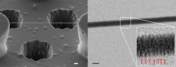
Obtained First DNA Image Through an Electron Microscope
 A team of researchers has succeeded for the first time to capture the image of DNA , the double helix model of DNA James Watson and Francis Crick proposed in 1953. DNA strands on a technique that will allow in the future to see how proteins, RNA and other biomolecules interact with DNA.
Crystallized atoms in DNA arrays to form a pattern of dots on a photographic film. The image interpretation requires complex math to figure out what the crystal structure could result in the observed patterns.
Now these new images are much more evident, as is direct images of the DNA strands , but views with electrons instead of X-ray photons How? The trick used by Enzo di Fabrizio, principal investigator of the University of Genoa, was DNA snag a dilute and put them on macroscopic silicon.
The team developed a model that is extremely pillars gua repellent, causing moisture to evaporate quickly leaving behind the DNA strands, which were stretched and could see clearly. Then, to obtain images of high resolution, tiny holes drilled on the basis of the silicon pillars.
Revealed results that the spiral thread of visible DNA double helix. One technique that scientists should be able to see individual DNA molecules in more detail and their interaction with proteins, RNA and other biomolecules.
Shortlink:

Recent Comments